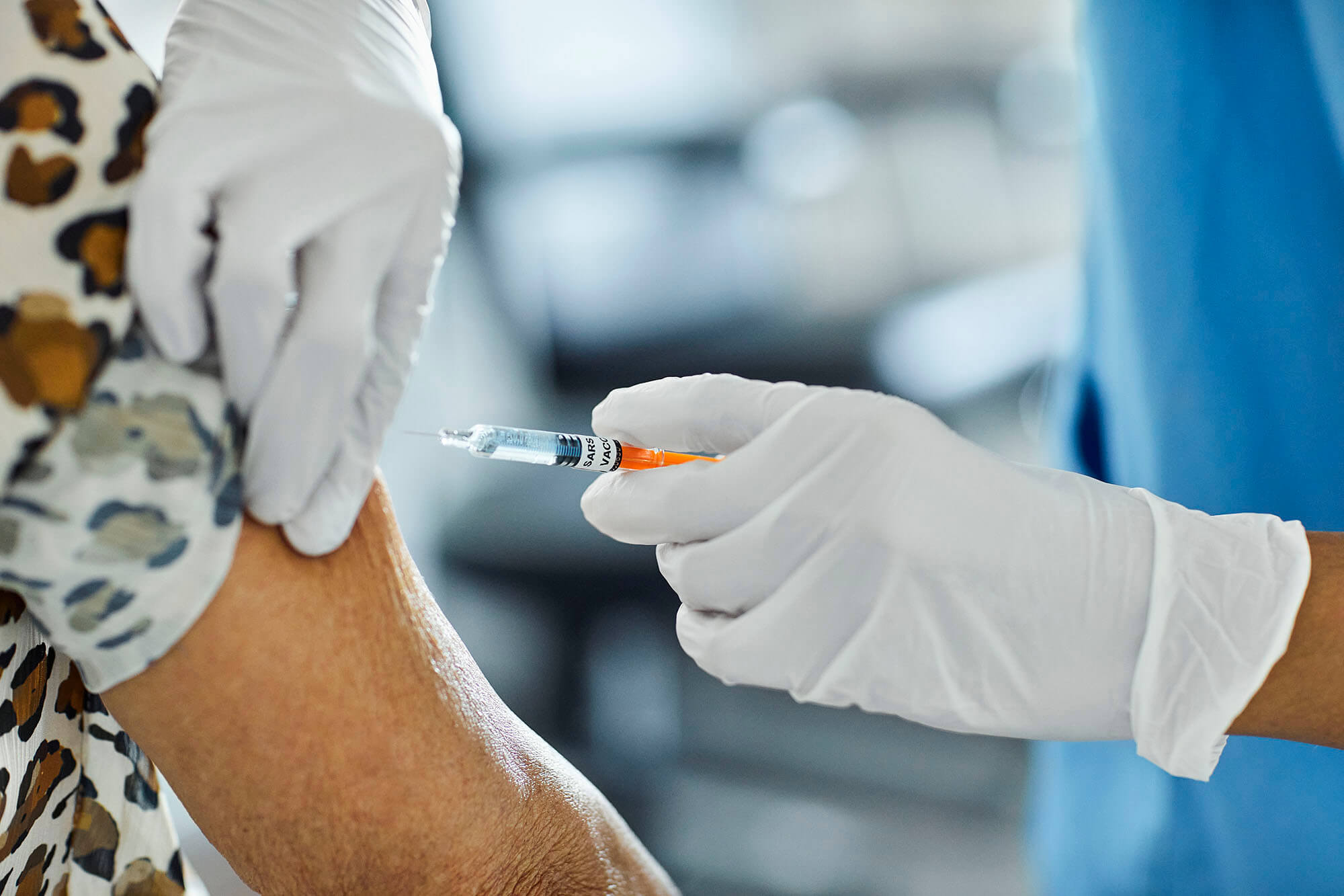
The United States is, according to a recent declaration from President Joe Biden, aiming to offer all US adults vaccination against SARS-CoV-2, the virus that causes COVID-19, by May 2021.1 To date, furthermore, more than 30 people per 100 across the country have received at least 1 dose of a currently approved vaccine.2
The vaccination program is likely to improve overall survival in the population of patients with cancer, given the high risk of COVID-19-related mortality in this group. The clinical considerations related to vaccination in this population are, however, not yet established, and some research suggests that oncologists and patients should be aware of the potential manifestations of vaccination on imaging and the consequences this might have on disease assessment, treatment monitoring, and decision-making.
A recent article published in the American Journal of Roentgenology suggests that vaccination can result in axillary, supraclavicular, and/or cervical lymph node enlargement on the side of the injection. Such enlargement may, according to the authors, lead to confounding results and/or even misinterpretation of fluorodeoxyglucose (FDG) positron emission tomographic/computer tomographic (PET/CT) imaging in some cases.
While lymphadenopathy has previously been reported with other vaccines, notably those against seasonal influenza and human papillomavirus, the widespread use of SARS-CoV-2 vaccination necessitates awareness in the clinical community of the potential for confounding findings on imaging, in particular with FDG PET/CT. Clinician awareness of vaccine site and timing can be useful with coordination of imaging to help avoid these manifestations that can, in some cases, interfere with the accuracy of the examinations. Yet given the number of patients with cancer who have received — or are likely to soon receive — vaccination, some scans may not be interpretable, leading to treatment indecision in the face of potentially aggressive disease.

To this end, Lacey McIntosh, DO, MPH, assistant professor at the University of Massachusetts Medical School in Worcester, and the study’s first author, discussed the implications of this research.
What is the dilemma with performing PET/CT in patients who have been recently vaccinated against COVID-19?
Dr McIntosh: The diagnostic dilemma that we are encountering is that the imaging manifestations of a recent COVID-19 vaccine can mimic cancer, especially on PET/CT. The vaccine causes tracer uptake and sometimes enlargement of lymph nodes of the axilla, supraclavicular region, and neck on the side of administration.
PET/CT is used for a variety of indications in oncology, including lesion characterization, disease staging, monitoring response to treatment, and surveillance for recurrence. Often, treatment decisions and disease status rely heavily on the results of this imaging. These studies can be difficult for physicians to get for their patients, so we want to make sure that when they are done, they are done well and give us the best chance to get the information we need with clear results.
In some cases, a recent vaccine just prior to imaging can potentially result in confusing findings rendering the study not useful and even lead to misinterpretation if information about vaccination is not available to the reading radiologist.
This article originally appeared on Cancer Therapy Advisor
this content first appear on medical bag

