
The severe systemic inflammatory processes and hypercoagulability occurring with COVID-19 illness increase the risk for atherosclerotic plaque disruption and acute myocardial infarction (AMI). Patients with a previous history of coronary disease and/or other significant comorbidities are particularly predisposed to cardiovascular complications with COVID-19 infection.1 In this installment, we will discuss a patient with COVID-19 and venous thromboembolism.
Case Presentation
A 61-year-old woman presents to a rural emergency department with complaints of progressively worsening dyspnea over the past 24 hours and pleuritic chest pain. On initial presentation, the patient is hypoxic with an oxygen saturation of 92% on 5 L/min supplemental oxygen via nasal cannula and exhibits sinus tachycardia (130-140 beats per minute).
The patient’s COVID-19 polymerase chain reaction (PCR) test is positive. Blood work reveals D-dimer is 3 times higher than normal (<0.4 mcg/mL), initial troponin within normal limits (0-0.1 ng/mL), hemoglobin 10.7 g/dL, hematocrit 33.1%, and platelet count 172 ×10/µL.
Continue Reading
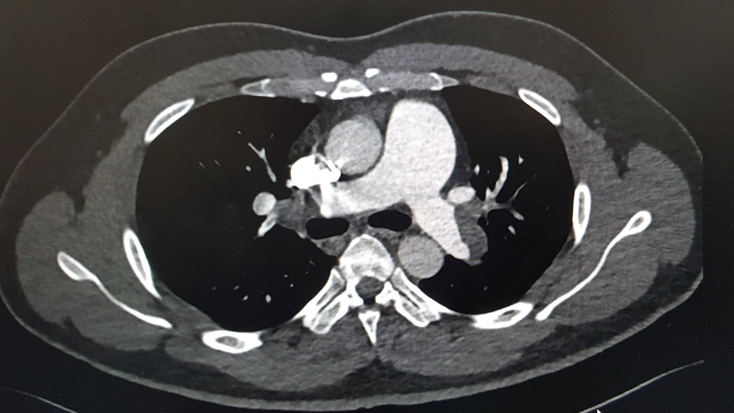
A massive saddle pulmonary emboli (PE) is present on spiral computed tomography (CT) arteriography with intravenous contrast of the pulmonary arteries. Echocardiogram demonstrates acute cor pulmonale with a right ventricular (RV) to left ventricular (LV) diameter ratio of 1.4. Venous ultrasound reveals a nonocclusive popliteal venous thromboembolism.
The patient is given full-dose enoxaparin and is transferred to an acute care cardiac specialty hospital for further treatment. Upon arrival at the specialty hospital, she is taken to the catheterization laboratory where a right and left pulmonary angiogram is performed with thrombectomy of the right and left pulmonary arteries.
Significant Medical History
The patient’s medical history includes type 2 diabetes mellitus, hypertension, dyslipidemia, hypothyroidism, and a 60-pack/year history of smoking.
Physical Examination
The patient is a middle-aged woman with obesity who is in acute respiratory distress. She has labored breathing and is tachypneic, with a respiratory rate in the mid-30s. Lung examination reveals mild expiratory wheezing bilaterally; a cardiac summation gallop is noted.
Electrocardiography (ECG) monitoring may indicate findings of cor pulmonale (right-sided heart failure) identified by a new incomplete or complete right bundle branch block, right axis deviation, or right ventricular ischemia with ST-segment depression in right pericardial leads. Monitoring with ECG also helps with evaluating for atrial arrhythmias such as atrial fibrillation commonly seen with PE.1
Spiral CT arteriography of the chest with contrast is ordered to rule out pulmonary embolus, which can be a contributing factor to respiratory symptoms, elevation in biomarkers, and a sequela of COVID-19 infection.3
Ultrasound of the lower extremities (bilaterally) is used to rule out deep vein thrombosis (DVT) in the lower extremities.
Diagnosis
The gold standard for confirmation of a PE is a spiral CT with arteriography. In this case, the test confirmed the presence of a massive saddle pulmonary embolus. Minimally invasive intervention is indicated if the patient is found to have right ventricular strain on echocardiogram (Table 1).
Table 1. Recommended Diagnostic/Laboratory Tests
| Coagulation: elevation in PT/INR, D-dimer, platelet count, fibrinogen |
| Cardiac biomarkers: troponin |
| Factor V Leiden mutation |
| Prothrombin gene mutation |
| Anticardiolipin antibodies (including lupus anticoagulant) |
| Hyperhomocysteinemia (usually due to folate deficiency)1,2 |
Radionuclide lung scan, commonly known as ventilation-perfusion (VQ) scan, may serve as a diagnostic tool for inpatients who have elevation in renal indices and are not able to undergo contrast studies. A VQ scan with a high clinical suspicion confirms the diagnosis of PE in 40% of cases.1
Echocardiogram is a useful tool for performing risk stratification. The presence of right ventricular wall akinesis or hypokinesis with sparing of the apex has a high specificity for acute PE. Also, in cases of PE, the ratio of the right ventricular end-diastolic area (RVEDA) to left ventricular end-diastolic area (LVEDA) exceeds the upper limit of normal, which is 0.6 mm.1
This article originally appeared on Clinical Advisor
this content first appear on medical bag